Showing posts with label cancer. Show all posts

Thursday, January 11, 2024

Did you know? Genetic Testing can help your pet better fight cancer!


Did you know...
DNA Genetic Testing is used to help pets with cancer!

Let’s delve into DNA genetic analysis! What exactly is it?

Genetic testing, also referred to as DNA testing, involves the use of technology or DNA sequencing to identify abnormal changes in genes, chromosomes or proteins. These changes, termed mutations or variants, often contribute to the formation and progression of cancer. DNA genetic testing is routinely used in human medicine and is anticipated to become a standard in veterinary oncology.

How can DNA genetic testing help my pet with cancer?

When DNA mutations or variants are identified through genetic testing, chemotherapy drugs that specifically target the identified mutations can be recommended as treatment for your pet’s cancer. Although DNA genetic testing is not perfect and may not always yield results, it can often help you and your veterinary oncologist make informed treatment decisions and can provide additional insights into prognosis for your pet. This precision allows veterinary oncologists to use precise and targeted chemotherapy to treat cancer, which decreases the risk of chemotherapy side effects.

DNA genetic testing can be especially helpful in challenging cases in which treatment options are limited. Ultimately, our goal is to extend your pet’s survival time past what is expected with conventional chemotherapy. By specifically targeting the mutations identified in your pet’s cancer, many times, targeted chemotherapy and conventional chemotherapy can be safely combined.

What samples are necessary for DNA genetic analysis?
Samples required for DNA genetic testing are typically obtained non-invasively. Depending on the specific test, blood, fine needle aspirates, or biopsy samples are necessary.

Is DNA genetic testing available for both dogs and cats?
Unfortunately, DNA genetic testing is exclusively available for dogs. However, there is hope that it will soon become accessible for cats.

My dog has been diagnosed with cancer. Is DNA genetic testing the next step?
Maybe! Not all cancers are treated equally, and chemotherapy is not always the chosen course of action. Sometimes, surgery alone might suffice for treating your pet’s cancer.

It is the goal of our Oncology Service is to help you explore the best treatment options for your pet and ensure treatment is focused on improving their quality of life. Learn more about our VCA Pet CancerCare Center and how our experts are here to care for your pet.


Dr. Irene Vazquez is a gifted and dedicated veterinarian who leads our Onolcogy department with a primary focus on improving quality of life for every pet that is diagnosed with cancer.
To schedule an appointment and to learn more about Dr. Vazquez, visit our website and watch this video.

Watch Video   


Advanced Cancer Care for the World's Most
Important Pet: Yours

At VCA, it is our goal to help treat your pet’s cancer, giving you and your pet more quality time together. At our VCA Pet CancerCare Centers, our oncology teams design individualized treatment plans for each patient using the most advanced and effective treatments available—much like those used in human medicine.

Learn about the VCA Pet CancerCare difference and watch the stories of pets we've helped after a cancer diagnosis.


Thursday, February 24, 2022

Roxy, A Canine Cancer Success Story!!!

Roxy is a 4 ½ year old female spayed small breed dog that presented to the Oncology Service for the first time on 4/21/21 for evaluation of a solitary right cranial pulmonary mass that was diagnosed on thoracic radiographs on 3/21/21 while she was being evaluated for coughing. An abdominal ultrasound was performed, which was normal, and cytology of the mass was performed and consistent with carcinoma. A chest CT was then performed with the goal of evaluating if Roxy was a good surgical candidate, but it unfortunately revealed multiple pulmonary nodules that were not visualized on radiographs. She was then diagnosed with metastatic pulmonary carcinoma and started on chemotherapy. 

Watch Roxy's story of concurring cancer!Roxy was treated with a combination of carboplatin and gemcitabine chemotherapy. She received a total of 8 cycles of chemotherapy and her total protocol duration time was 6 months. She did not experience any side effects from chemotherapy and her quality of life was excellent while undergoing treatment. She had thoracic radiographs performed prior to cycles #4 and #8, and both showed no evidence of cancer, which means she entered complete remission with chemotherapy. She was then transitioned to maintenance oral chemotherapy and was still in complete remission on 10/13/21 when she had radiographs last performed. Thanks to chemotherapy, she has been alive for 7 months after a diagnosis of metastatic pulmonary cancer. Without chemotherapy, her expected survival time was less than 3 months.

In summary, dogs with pulmonary carcinoma can have positive outcomes if treated appropriately and if no negative prognostic factors are identified. The presence of one or more identified negative prognostic factors can negatively affect prognosis. Although surgery is the mainstay treatment for this type of cancer, the addition of chemotherapy in efforts to prolong survival time in pets with identified negative prognostic factors can be helpful. For dogs that are not good surgical candidates, chemotherapy is a palliative option that can be pursued as in Roxy’s case.

A Case of Canine Metastatic Pulmonary
Carcinoma


Irene.Vazquez@vca.com
Oncology Services
Monday-Thursday


References

1. Rebhun, R.B., Culp, W.T.N., (2007). Pulmonary Neoplasia. In Withrow & MacEwen's Small Animal Clinical Oncology (5th ed., p. 453-459). St. Louis, MO: Saunders Elsevier.

2. Brody RS, Craig PH: Primary pulmonary neoplasms in the dog: a review of 29 cases, J Am Vet Med Assoc 147:1628-1643, 1965.

3. Griffery SM, Kraegel SA, Madewell BR: Rapid detection of K-ras gene mutations in canine lung cancer using single-strand conformational polymorphism analysis, Carcinogenesis 19:959-963, 1998.

4. Meuten DJ: Tumors in domestic animals, ed 4, Ames, Iowa, 2002, Iowa State University Press.

5. Hahn FF, Muggenburg BA. Griffith WC: Primary lung neoplasia in a beagle colony, Vet Pathol 33:633-638, 1996.

6. Moulton JE, von Tscharner C, Schneider R: Classification of lung carcinomas in the dog and cat, Vet Pathol 18:513-528, 1981.

7. Nielsen SW, Horava A: Primary pulmonary tumors of the dog. A report of sixteen cases, Am J Vet Res 21:813-830, 1960.

8. D’Costa S, Yoon B-I, Kim DY, et al: Morphologic and molecular analysis of 39 spontaneous feline pulmonary carcinomas, Vet Pathol DOI 10.117710300985811419529, 2011.

Friday, September 3, 2021

Mast Cell Tumors Beware! Our Oncologist has a mark on you!

Mast Cell Tumors Beware! Our Oncologist has a mark on you!   


When looking for advanced veterinary care, we are here for you with cutting edge treatment options. Dr. Irene Vazquez, our board certified oncologist is your neighborhood pet cancer specialist.

STELFONTA is a prescription medicine used in dogs to treat mast cell tumors on the skin or in the tissues just under the skin. Given as an injection directly into your dog’s tumor, it destroys the tumor by breaking down cancer cells and starving their blood supply. A “pocket” or wound is left at the injection site where the tumor was. This is generally a healthy wound ready for skin and hair to grow back in a few weeks.
Pretreatment involves oral medications, including corticosteroids and antihistamines, prior to the injection of STELFONTA. These medications are very important to reduce the risks associated with inflammatory substances that can be released from the tumor as the mast cells are destroyed. Premedication is given 2 days pretreatment and continues until 7 days post treatment. It is essential that you give all medications as prescribed.

Most dogs do not need sedation or anesthesia during the procedure although occasionally, dogs may be sedated.

Side effects reported in dogs treated with STELFONTA were generally mild and short lasting, but they can be severe, especially if not treated in a timely manner.

The most common adverse reactions included wound formation, injection-site pain, lameness in the treated limb, vomiting, diarrhea and hypoalbuminemia (low levels in the blood of the protein albumin). Although most side effects/adverse reactions seen in clinical studies were mild, the most serious side effects occurred in the first 4 days. Excessive pain, lameness, tiredness, refusal to eat for more than one day, repeated vomiting or diarrhea, trouble breathing, changes to the treated tumor site (including increased or excessive swelling and bruising, extensive wound formation, increased irritation) or any other symptoms should be examined immediately and treated accordingly.

When looking for advanced veterinary care, we are here for you with cutting edge treatment options. Dr. Irene Vazquez, our board certified oncologist is your neighborhood pet cancer specialist.

Irene Vazquez, DVM, MS, DACVIM (Oncology)


Thursday, September 2, 2021

New Treatment for Mast Cell Tumors (MCT)-STELFONTA

New Treatment for Mast Cell Tumors (MCT)-STELFONTA

VCA Hollywood Animal Hospital now offers STELFONTA as a cutting edge treatment for Mast Cell Tumors (MCT). STELFONTA is a prescription medicine used in dogs to treat mast cell tumors on the skin or in the tissues just under the skin. Given as an injection directly into your dog’s tumor, it destroys the tumor by breaking down cancer cells and starving their blood supply. A “pocket” or wound is left at the injection site where the tumor was. This is generally a healthy wound ready for skin and hair to grow back in a few weeks.

Pretreatment involves oral medications, including corticosteroids and antihistamines, prior to the injection of STELFONTA. These medications are very important to reduce the risks associated with inflammatory substances that can be released from the tumor as the mast cells are destroyed. Premedication is given 2 days pretreatment and continues until 7 days post treatment. It is essential that you give all medications as prescribed.

Most dogs do not need sedation or anesthesia during the procedure although occasionally, dogs may be sedated. 

Side effects reported in dogs treated with STELFONTA were generally mild and short lasting, but they can be severe, especially if not treated in a timely manner. 

The most common adverse reactions included wound formation, injection-site pain, lameness in the treated limb, vomiting, diarrhea, and hypoalbuminemia (low levels in the blood of the protein albumin). Although most side effects/adverse reactions seen in clinical studies were mild, the most serious side effects occurred in the first 4 days. Excessive pain, lameness, tiredness, refusal to eat for more than one day, repeated vomiting or diarrhea, trouble breathing, changes to the treated tumor site (including increased or excessive swelling and bruising, extensive wound formation, increased irritation) or any other symptoms should be examined immediately and treated accordingly. 


Medical Oncologist
VCA Hollywood Animal Hospital
vcahollywood@vca.com


Friday, August 27, 2021

Message from our Oncology Department:

Welcome back, Dr. Vazquez!!!!!!

We are pleased to announce that Dr. Irene Vazquez, returns from maternity leave on Tuesday, September 7th to resume her full schedule of appointments and procedures Monday- Thursday.

 We are very excited to have her back and look forward to sharing with you some of the newest treatments she is implementing in her cancer care service.

As always our Emergency Service is open
24/7/365.
Our Specialized Veterinary Care is available
Monday-Saturday!

We understand the unique impact this year has had on all of us and we are grateful to be your source for emergency & specialty pet care.

Learn more about Pet Cancer Care and the vast resources that are available through your local VCA Hollywood Pet Cancer Care Center.

Tuesday, February 25, 2020

We Can-cer-vive Cancer! Meet Charlie!!!

Meet cancer survivor Charlie and learn how a team approach to health care is so important when it comes to your pet's health. 

Charlie is a very adorable 14 year old cat that was diagnosed with an ear canal cancer known as ceruminous gland adenocarcinoma. This type of cancer is one of the most common ear canal cancers seen in both cats and dogs. Clinical signs associated with this disease include excessive cerumen accumulation, discharge, swelling, itchiness, and recurrent infections. In order to obtain a diagnosis, advanced imaging such as a CT scan is usually required. 

Most commonly, ear carcinomas are locally invasive and have a low chance of spreading to other locations. The treatment of choice for this type of cancer is surgical resection, which involves completely removing the ear canal (TECA- total ear canal ablation). 


A TECA or in Charlie's case a TECABO is a Total Ear Canal Ablation or Total Ear Canal Ablation Bulla Osteotomy. This procedure involves the complete removal of the ear canal middle ear, leaving only the ear flap remaining. 


Although this type of surgery may sound like a lot for your pet to go through, they tend to feel so much better once it is performed and it gives them the best chance at achieving a long survival time. With successful surgery and no identified negative prognostic indicators, patients can live a long time after removal of their cancer. 

Charlie was initially evaluated by Dr. Irene Vazquez at our the Oncology Service and had a CT scan performed, which showed he had a mass within his ear canal. The mass was sampled and the results were consistent with carcinoma. Charlie was then evaluated by Dr. Andrea Clark at our Surgery Service and had surgery performed to remove his tumor. 

Charlie on his 5 month anniversary cancer free!
Since his surgery, Charlie has been doing great at home and is currently cancer free. He continues to come in for routine evaluations to make sure his cancer is not spreading and so far, he's been doing amazing! He is the sweetest boy and our hearts melt every time he comes in. 

We wish Charlie the best of luck and the longest survival time! Our VCA Hollywood family is SO proud of him ðŸ’™.


Contributors: Irene Vazquez, DVM and Andrea Clark, DVM
Charlie after surgery. 


Ear Canal Tumors

What are tumors of the ear canal?
Tumors of the ear canal are abnormal growths that can develop from any part of the ear canal (the skin, the glands of the skin that produce earwax and oil, and the underlying connective tissues, muscles, and bones). Ear canal tumors can be benign or malignant. Benign tumors do not spread, while malignant or cancerous tumors can spread to the surrounding tissues or even other areas of the body (metastasis). Tumors of the external ear canal are more common than tumors of the middle or inner ear.

In dogs, the most common external ear canal tumors are ceruminous gland adenomas (benign) and adenocarcinomas (malignant). Other tumors include inflammatory polyps, papillomas, sebaceous gland adenomas, and more.

In cats, the most common external ear canal tumors are nasopharyngeal polyps, squamous cell carcinomas, and ceruminous gland adenocarcinomas.

Lymphoma, fibrosarcoma, and squamous cell carninomas are occasionally seen in the middle or inner ear of dogs and cats.

What causes these kinds of tumors?
The reason why a particular pet may develop this, or any other tumor or cancer, is not straightforward. Very few tumors and cancers have a single known cause. Most seem to be caused by a complex mix of risk factors, some environmental and some genetic or hereditary. Although there does not appear to be a genetic or hereditary cause for ear canal tumors, chronic inflammation is believed to play a role. German Shepherds and Cocker Spaniels, for example, are at increased risk of developing these tumors. This may be related to the tendency of both breeds to have recurrent and severe ear infections.

What are the clinical signs of ear canal tumors?
horners_syndrome_dog_arrow_2018Initially, these types of tumors may appear as one or more pink, white, or purple nodular masses in the ear canal. If benign, they may grow to a certain size and may or may not be problematic. If malignant, they may grow, ulcerate (break open) and bleed, and nearly invariably become infected, causing recurrent or chronic ear infections. As they grow, they can narrow and even fill (and obstruct) the ear canal.

The most common clinical signs of pets with ear canal tumors include an inflamed, itchy and painful ear, persistent odorous discharge (that can be waxy, pus-filled, or bloody), head shaking, and ear scratching. Sometimes pets will shake or scratch hard enough to cause an ear hematoma (a blood blister). Occasionally draining abscesses may form just below the ear. If the middle or inner ear is involved, pets may experience head tilt, circling, loss of balance and coordination, nystagmus (darting of the eyes back-and-forth), Horner’s syndrome (see photo), facial paralysis (with facial drooping, salivation, and difficulty eating), loss of hearing, and other neurologic signs.
(Image via Wikimedia Commons / Joel Mills (CC BY-SA 3.0.)

How are these kinds of tumor diagnosed?
If deep in the ear canal, these tumors may be difficult to see and therefore diagnose. If the ear canal is severely inflamed, the inflammation may need to be alleviated first to more clearly see the mass. Once the mass is identified, it may (or may not) be possible to take a sample of the tissue, depending on the location. If the mass can be accessed, fine needle aspiration (FNA) or biopsy will be performed. FNA involves taking a small needle with a syringe and suctioning a sample of cells directly from the tumor and placing them on a microscope slide. A veterinary pathologist then examines the slide under a microscope. A biopsy is surgical removal of a piece of the tumor. The tumor tissue is then examined under the microscope. This is called histopathology.

If a diagnosis of malignancy is made from the FNA or biopsy, a CT scan of the head and neck region may be performed to determine how invasive the tumor is and plan a surgical approach. Staging (searching for potential spread to other locations in the body) is highly recommended for malignant tumors, as they tend to spread. This may include bloodwork, urinalysis, X-rays of the lungs, and possibly an abdominal ultrasound. If any lymph nodes appear to be enlarged or firmer than normal, samples may be taken to look for spread.

How do these types of tumor typically progress?
Benign tumors, such as inflammatory polyps, typically only grow to a certain size, but are often irritating and interfere with the ear’s normal function, resulting in waxy buildup and chronic infections. The biggest concern with malignant tumors (other than the possibility of metastasis) is local spread. If the tumor is not treated, it may continue to grow and invade the surrounding tissues, causing pain and increasing the risk of the more serious clinical signs and complications listed above.

What are the treatments for these types of tumor?
The treatment of choice for ear canal tumors is surgical excision. This is curative for benign tumors that are completely removed. Laser surgery may be an option. More extensive surgery is required for malignant ear canal tumors. One procedure, called total ear canal ablation and bulla osteotomy (TECA-BO), involves removing the outer and inner ear canal, as well as the tympanic bulla (the middle ear), leaving only the ear flap remaining.

If complete removal of the tumor is not possible, radiation therapy may be recommended to treat the remaining tumor cells. Radiation therapy may also be recommended for large tumors that cannot be surgically removed, to slow tumor growth or relieve pain. Chemotherapy may be an option if the tumor is highly malignant or if there is evidence of metastasis.

Is there anything else I should know?
It is important to recognize that tumors of the ear canal may grow without much outward evidence, only causing subtle signs in your pet. If your pet suffers from recurrent or chronic ear infections, or shows signs of pain or discomfort, rechecking with your veterinarian at frequent intervals may assist with early detection.

Contributors: Christopher Pinard, DVM
© Copyright 2018 LifeLearn Inc. Used and/or modified with permission under license.
https://vcahospitals.com/know-your-pet/ear-canal-tumors

A Team Approach to Cancer Care, VCA Pet Cancer Care Center!

A diagnosis of cancer is stressful for every pet owner. Helping owners learn as much as they can about the disease and the latest advances in treatment is key to managing fear, confusion, and helping them make an educated decision on their pet’s health.

Our mission is to provide an exceptional level of progressive and sensitive cancer care to the cancer patient through a team approach and ensure that our client is always well informed and comfortable with decisions made regarding their pet’s care.

Using the strengths and expertise of all specialists on site, including doctors from our Surgery, Internal Medicine, Cardiology, Dermatology, Radiology, and Emergency and Critical Care departments we tailor treatment protocols for each pet’s specific cancer and physical conditions to optimize the outcome and minimize side effects.

Our technicians have experience in caring and supporting cancer patients. They are knowledgeable and experienced in chemotherapy administration and treatment and they adhere to strict protocols to maintain the highest level of safety for the staff and patient. We emphasize support, education, and improving the quality of life for all animals, particularly those undergoing treatment.

Our Medical Oncology department utilizes chemotherapy to treat cancer.  Each tumor type has a different biological behavior. Depending on the tumor type, staging exams are usually performed prior to recommending treatment. Staging exams may include fine needle aspirates and/or biopsies, chest radiographs, an abdominal ultrasound and ultrasound guided aspirates, CT scan, etc. These can all be performed on-site and help us gain a better idea of the pet’s overall health.  Based on the tumor type and staging exam results, we may recommend surgery, chemotherapy, or a combination of both treatments. For some types of cancer, a curative intent treatment can be pursued. For others, a palliative intent treatment may be followed. 
 VCA Hollywood offers Medical Oncology Services
 with Dr. Irene Vazquez.
 Get to know Dr. Vazquez!

Thursday, January 30, 2020

The Five Most Common Questions about Cancer in Dogs

How common is cancer in dogs? Unfortunately, cancer is one of the leading causes of death in older animals. According to the Animal Cancer Foundation (ACF), 1 out of every 4 dogs will develop cancer in their lifetime, and each year, approximately 6 million dogs are diagnosed with cancer.

What are some of the most common cancers in dogs?
Like their human companions, dogs can develop many different types of cancer, such as mammary cancers, lymphoma, leukemia, osteosarcomas, mast cell tumors and skin cancers, to name a few. According to the Veterinary Cancer Society, mammary tumors are the most common cancer seen in dogs.

Are there diagnostic tests or exams that should be done regularly to try to detect cancer in dogs? 

To improve the chances of early cancer detection, you should take your pet to your veterinarian at least once a year. Personally, I recommend animals over 6 years of age be seen twice a year. Your veterinarian will do a complete examination, checking your dog from head to tail to look for:

  • Abnormal skin growths (lumps and bumps)
  • Enlarged organs or masses in your pet’s abdomen (belly)
  • Abnormal heart or lung sounds, and abnormalities in the eyes, ears and mouth
These exams are important because veterinarians can discover things that you may never notice. However, not all cancers can be detected by physical examination, so your veterinarian may also recommend screening tests like blood work, urinalysis and radiographs (x-rays), to uncover underlying diseases like cancer. Depending on the age, sex and breed of your dog, your veterinarian may recommend one or all of these tests to help look for cancer.

Are some types of cancers more treatable than others? 
Yes, all cancers are different and some are more treatable than others. The type of cancer, location, size, stage and response to treatment all affect the ultimate prognosis. With many cancers, the earlier the treatment is started, the better the prognosis, and ultimately, the longer the survival times. That's why it is so important to bring your dog to the veterinarian for examinations at least once a year. 

Do chemotherapy and radiation treatments affect dogs the way they affect human cancer patients? 

While remission is the ultimate goal, quality of life is equally as important. As a result, cancer treatments in dogs are generally less aggressive than regimens used in people. Therefore, pets typically do not suffer from some of the troubling side effects seen in people, such as nausea, vomitinghair loss and extreme fatigue. If side effects occur, your veterinarian will adjust the treatment to ensure that your pet remains as comfortable as possible.

The goal of cancer therapy in dogs, like with many other diseases, is early diagnosis and treatment. In order to give your dogs the best odds of beating cancer, make sure your dog gets regular check-ups, take advantage of recommended screening tests, and become familiar with the common signs and symptoms of cancer in pets so you can look-out for them at home. If you want to know more about cancer, speak with your veterinarian and visit Veterinary Cancer Society or Animal Cancer Foundation 

If you have any questions or concerns, you should always visit or call your veterinarian -- they are your best resource to ensure the health and well-being of your pets.


Wednesday, January 15, 2020

We Can-cer-vive Cancer! - #VCAHollywoodPetCancerCareCenters


Oscar is a 10 year old Shih Tzu that was diagnosed with intestinal lymphoma on September 23, 2019.


Lymphoma is a type of cancer that originates from the abnormal growth of lymphocytes, an important cell of the immune system. Lymphoma in dogs most commonly affects the lymph nodes, but it can also affect the lymphatics within the intestinal tract. Although intestinal lymphoma is not a disease that can be cured, treatment with chemotherapy can help keep the cancer at bay for a period of time, prolong their lifetime, and most importantly, greatly improve their quality of life.


Oscar underwent 15 weeks of chemotherapy with the *CHOP protocol and received his last chemotherapy treatment today! He did great throughout his chemotherapy protocol and experienced minimal to no side effects.

He's our little trooper and a chemo rock star! We are so proud of him and will miss seeing him through our Oncology Service.  We wish he and his family the best of luck!


VCA Hollywood's Oncology Department applies a team approach to cancer care using the strength & expertise of all of our on site specialists Surgery, Internal Medicine, Cardiology, Dermatology, Radiology and Emergency & Critical Care departments to provide the very best care


Dr. Vazquez joined our team in July of 2019 after completing her Oncology Residency at Auburn Univeristy. For Dr. Vazquez, the most important thing in dogs and cats after a diagnosis of cancer is that  treatment is focused on improvement of their quality of life.


* CHOP is the 'gold standard' of  treatment for dogs with multicentric lymphoma consists of a multi-agent, CHOP-based chemotherapy protocol incorporating several injectable and oral drugs given on a more-or-less weekly basis for a period of several months.


Tuesday, May 17, 2016

Patient highlight- Jade!!!

 This is Jade. Jade is a sweet girl who unfortunately had a tumor in her left tibia. The type of tumor she had is known as osteosarcoma.

Osteosarcoma is a primary tumor of bone meaning, that the tumor arises from the cells that make up bone. Bone cancer is not that common in cats however,  of the tumors of bone found in cats, osteosarcoma is the most common type.

Poor Ms. Jade had a tumor that affected the leg so much that the bone broke.  The treatment was, amputation of the leg. The prognosis often depends upon species (dogs often have a different prognosis than cats) and how aggressive the tumor is.

Jade had an amputation with one of the board certified surgeons at VCA Hollywood Animal Hospital, Dr. Briana Danielson.

Jade did very well with her surgery and while it did take a little while for her to figure out how to get around on three legs, she's doing great now and feels good!

We are hopeful that she will continue to do well and live a very long life. We are so happy for Jade and her family.

It's she so cute rolling on her back to show us her belly. What a beautiful girl she is!


Thursday, December 26, 2013

Breast Cancer Awareness!

Breast Cancer in Pets. (yes, they get it too!!)


Many pet parents don't realize that pets can also suffer from breast cancer. In veterinary medicine, these tumors are referred to as mammary gland tumors, and are unfortunately one of the most common kinds of cancer in pets.

Cats generally have eight mammary glands, arranged in four pairs. Dogs usually have 10 glands arranged in five pairs, though the number varies with the size of the dog. Mammary gland tumors in dogs and cats can be benign or malignant. In cats, around 90% of mammary gland tumors are malignant. In dogs, approximately 50% are malignant.

How can mammary gland tumors be prevented in dogs and cats?
The most effective way to prevent mammary gland tumors is to have your pet spayed before she ever goes into heat. There is a myth that animals should have one heat cycle (or give birth to one litter) before they are spayed. In fact, dogs who are spayed before their first heat cycle are 2,000 times less likely to develop breast cancer! Cats spayed before their first heat are 91% less likely to develop breast cancer than unspayed cats. After just one heat cycle, the risk rises in both dogs and cats.

Detecting mammary gland tumors
Just like in people, performing mammary exams in dogs and cats is very important. Early detection is key. If your dog or cat allows, perform a mammary exam on her once a month. Gently feel the tissue under and around each nipple, "rolling" the tissue between your fingers. Very small mammary tumors often feel like a little BB pellet under the skin. If you feel even a tiny lump or firm area, bring your pet to the veterinarian immediately.
There is evidence that canine mammary tumors can become malignant over time, so prompt removal is essential.

Treatment of mammary gland tumors in pets
The main treatment at this time is surgical removal. Depending on the situation, your pet may need to have the affected mammary gland, several mammary glands, or all the glands on that side of her body removed. The tumor that is removed will be sent to the lab for a biopsy to tell you if it is benign or malignant. If the tumor is malignant, your veterinarian may refer you to a veterinary oncologist for consultation.
For more information about keeping your pet healthy, please visit the ASPCA Animal Hospital.


Source:  http://www.aspca.org/blog/term/pet-care
Friday, December 13, 2013 - 3:45pm
Guest blog by Louise Murray, DVM DACVIM, Vice President of the ASPCA Animal Hospital and author of “Vet Confidential: An Insider’s Guide to Protecting Your Pet’s Health”

Monday, January 14, 2013

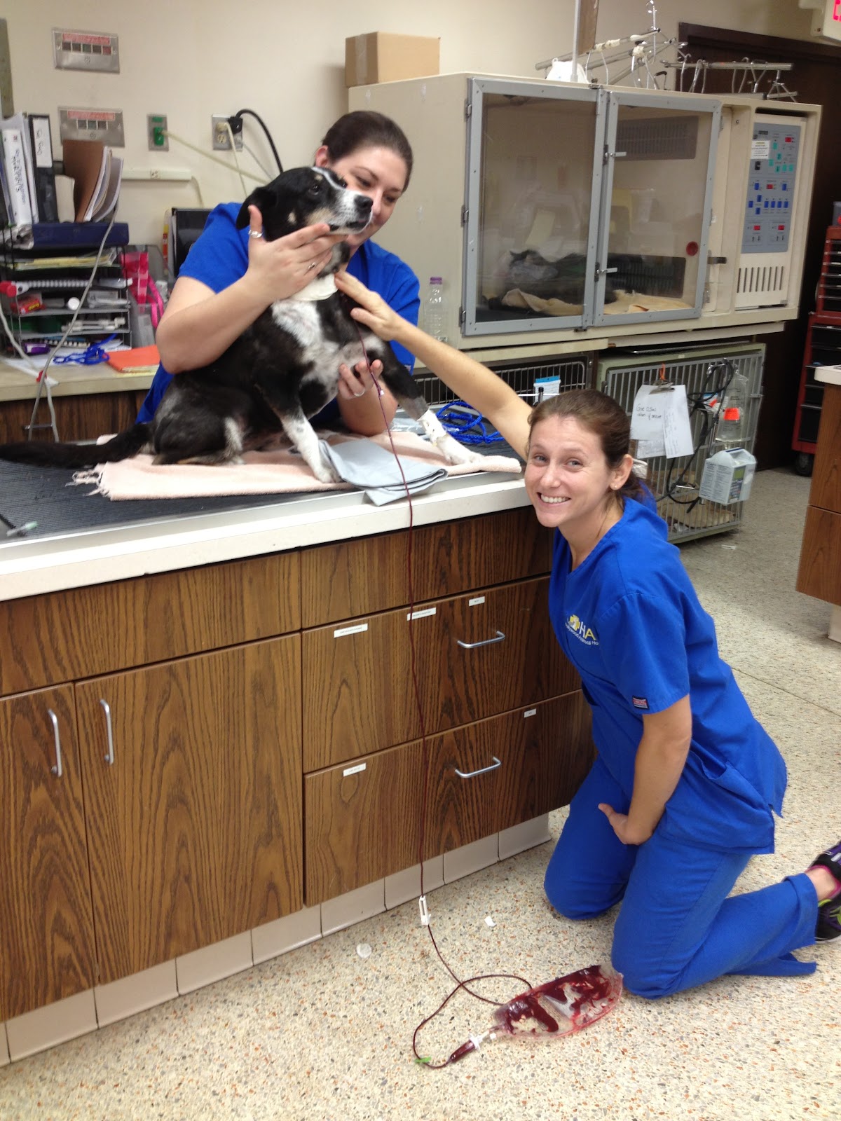
January- Patient of the Month "Sarah"


Sarah, a 10year old, collie mix has been a patient of the HAH since 2006. From annual exams to a mass removal in 2009 her history of care was typical.
However, in 2011, Sarah collapsed and was brought to the HAH for evaluation. Blood work showed abnormalities only in her PCV (packed cell volume). The nature of her presentation and this abnormal blood level alarmed Dr. Diamant and she discussed the possible of a Polycythemia Vera diagnosis. To confirm, Dr. Diamant ordered an ultrasound and bone marrow biopsy.
Polycythemia vera is a bone marrow cancer mainly causing the production of too many red blood cells.  Some of the changes associated with Polycythemia vera are abnormal motor skills, brick red gums, pale gums, nose bleeds and an enlarged spleen.

Once the diagnosis was confirmed, Sarah’s owners were faced with treatment options. This kind of blood cancer is treated in primarily two ways; chemotherapy and phlebotomy.

The chemotherapy for polycythemia versa is costly and Sarah’s owners opted for phlebotomy first. Phlebotomy is used to decrease blood thickness. One unit of blood (about 1 pint) is removed weekly until the hematocrit level (PCV) is less than 45. Therapy is continued as needed.

Currently, Sarah is under the care of Dr. Carlos and comes in approximately every three weeks and has blood drawn two times. Each time about 300mls is removed and her PCV is checked. Sarah is a wonderful patient. She seems unaffected by the blood draws and loves the extra attention she gets.

Sarah is regular at the HAH and always receives a regulars welcome. We are so pleased with her progress and anticipate continued success. We are pleased to honor her as patient of the month!